Technology

Intra Oral Camera
Intra Oral Cameras allow dentists to capture and display digital images of the oral cavity. High resolution images captured by the intra oral camera are displayed on a chairside monitor. These images can aid in diagnosing surface lesions as well as increase transparency in your treatment planning by giving you a clearer understanding of what the dentist sees. These images can also indicate areas vulnerable to plaque build up or cavities. This can increase the quality of your at home care by educating you on your dental and gingival architecture. reducing the need for more invasive procedures in the future.
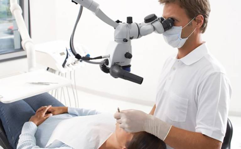
Dental Microscope
Dental Operating Microscopes, or dental surgical microscopes, are designed to provide ideal magnification of the area of the mouth being worked on while also allowing the clinician to maintain an ergonomic working position. This enables the dentist to maintain a comfortable position, obtain an optimal view of the working area, and focus on the patient. Most dental microscopes are controlled via foot pedal for hands-free operation. While microscopes are most often used during endodontic procedures, the enhanced magnification they provide can be ideal for use during oral surgery, laser dentistry, restorative procedures, and a range of other clinical situations. In addition to the basic microscope, there are also additional options available. Binocular heads allow another dentist, tech or student to simultaneously view a surgery, while a camera attachment allows the images to be streamed to a monitor so an audience can view the surgery, providing a valuable teaching tool. These videos also can be recorder to allow for documentation of treatments. Installation options include wall mounting or a portable stand.

Cone Beam Computer Tomography (CBCT) Scanner
CBCT scanner creates a 3 dimensional model of your mouth by stitching multiple images together. The scanner will rotate around your head taking multiple x-ray images. These images are then compiled into a single 3D model. Through this model, the doctor is able to visualize the locations of the teeth and the surrounding vascular and hard tissue. Any dental abnormalities, canals, subsurface lesions, nerves, and vessels can be seen. This allows the dentist to more precisely plan their approach and prevents iatrogenic errors from occurring, minimizing risk of complications. Because of these benefits, CBCTs have become the standard in ensuring safety and effective practice during endodontic and surgical procedures.

iTero® digital scanning
The iTero® digital scanning system eliminates the need for traditional dental impressions. Whereas traditional dental impressions are both uncomfortable to take and inaccurate when casted, 3D digital scanning is not only faster, but provides higher resolution and more accuracy. Additionally, the software can fabricate digital models of crowns and bridges and can even simulate the results of orthodontic treatment, giving you a better understanding of your treatment plan. Digital dentistry reduces chairtime by minimizing fabrication errors and decreasing the amount of appointments needed, meaning that you can receive your dental appliances quickly and comfortably.

Surgical laser WaterLase® iPlus
Laser dentistry has been the newest development in the field, bringing faster and more comfortable delivery of dental care. As we prioritize your comfort and satisfaction, our office has adopted the WaterLase dental laser. Laser dentistry is applicable in procedures such as gum contouring, decay removal, bone surgery, and cosmetic procedures. Waterlase uses a water stream and laser energy to cut hard and soft tissue, which avoids creating heat, making Waterlase virtually painless. This not only removes the need for anesthesia, but also reduces post-op sensitivity and pain. In comparison to traditional handpieces, Waterlase provides more precise removal of decay and incision of tissue, increasing the safety and efficacy of procedures.

Digital X ray sensor
Our practice utilizes digital x-ray images. In comparison to traditional film based x-rays, CCD digital x-ray sensors require less exposure time, reducing your amount of radiation exposure. Digital x-ray sensors also transmit the radiograph to a computer instantly and display it at a higher resolution than film x-rays. The viewing program also has the ability to digitally alter the x-ray’s characteristics, increasing visibility of any potential lesions, resulting in a more accurate diagnosis and earlier treatment.